
Examples SeeNano Lab
Examples > SeeNano Lab
With the Grayfield microscopes, there is no interdependance between depth of field, magnification and resolution. Depth of field is adjustable between "very large" and "none" allowing for an image depth that was previously not possible.
Click on the images below to enlarge...
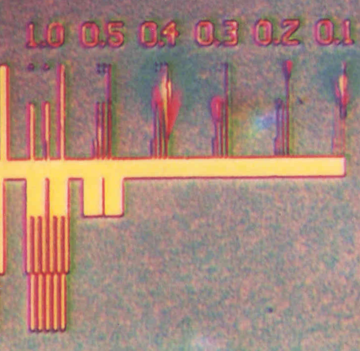
Test Chip. An overview from 1000nm to 100nm
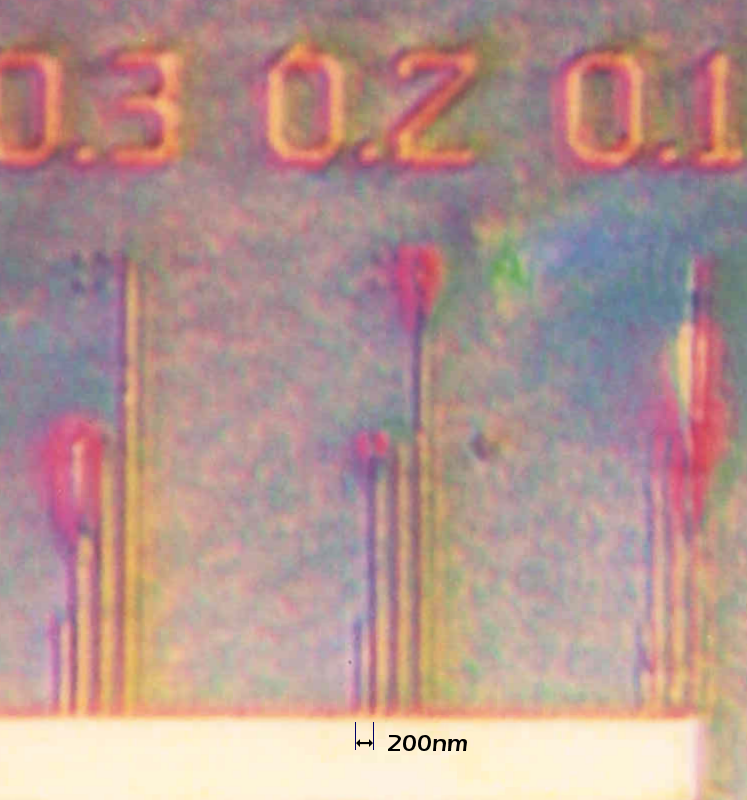
Shows sharp images at 300 nm / 200 nm, and detectable at 100 nm.
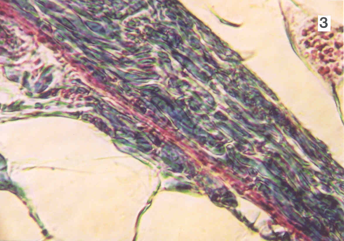
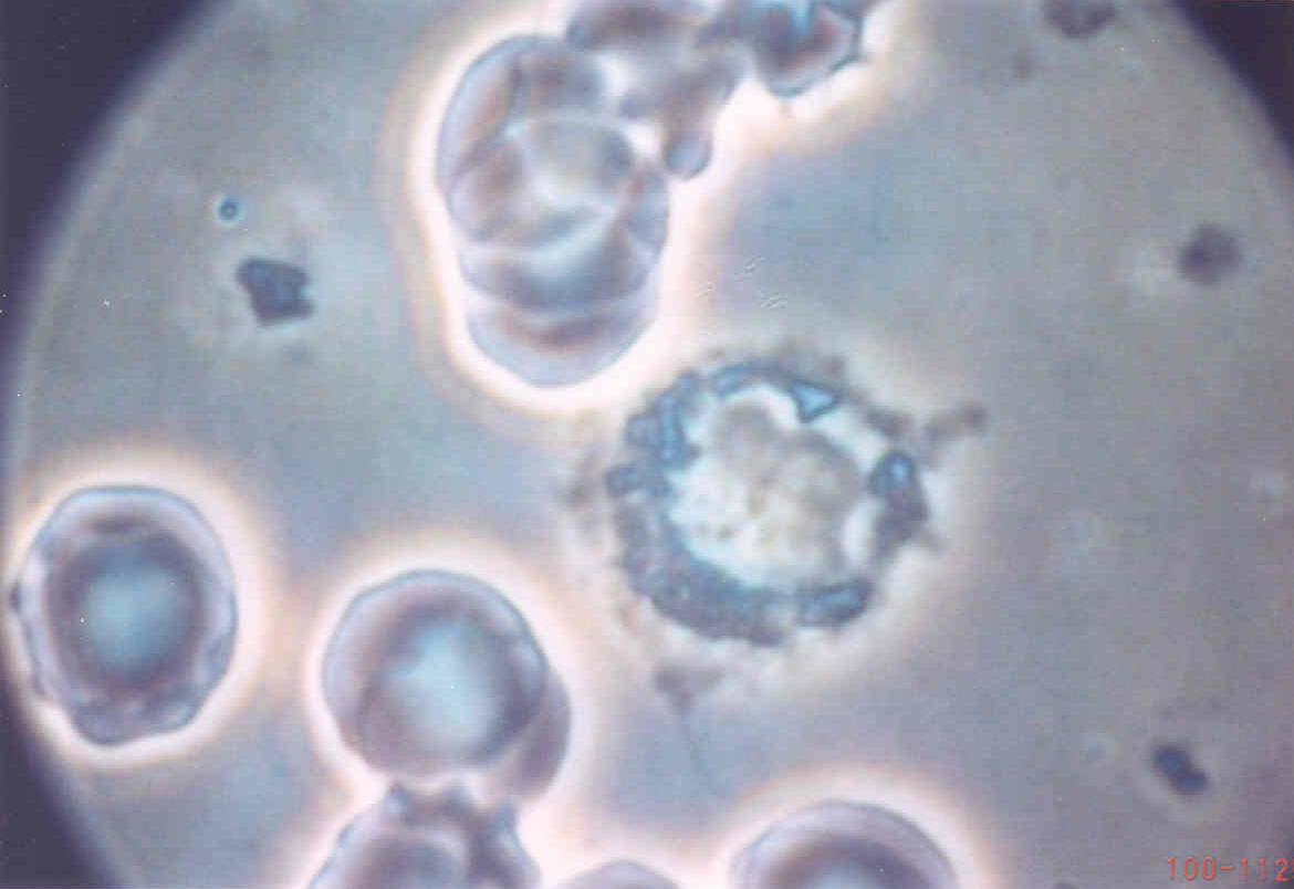
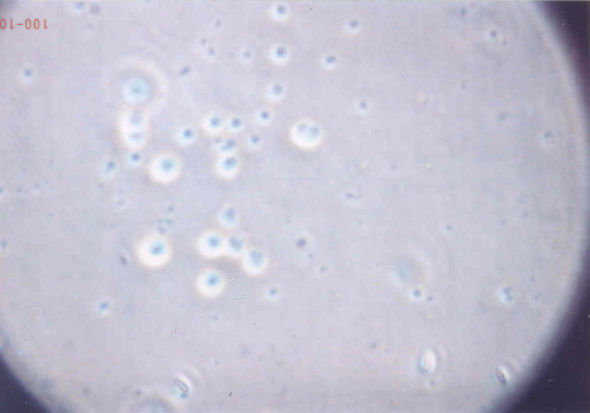
Tissue sample - 20x Phase contrast
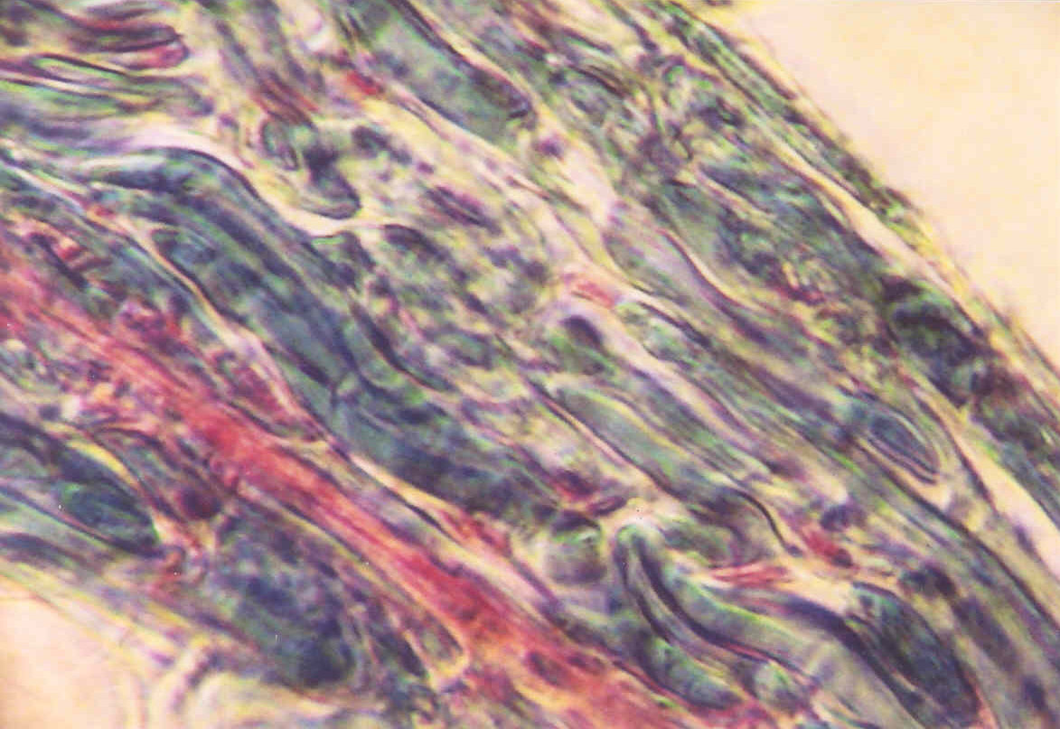
Tissue sample - 40x Phase contrast
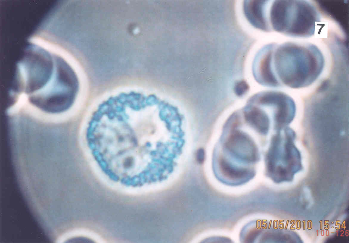
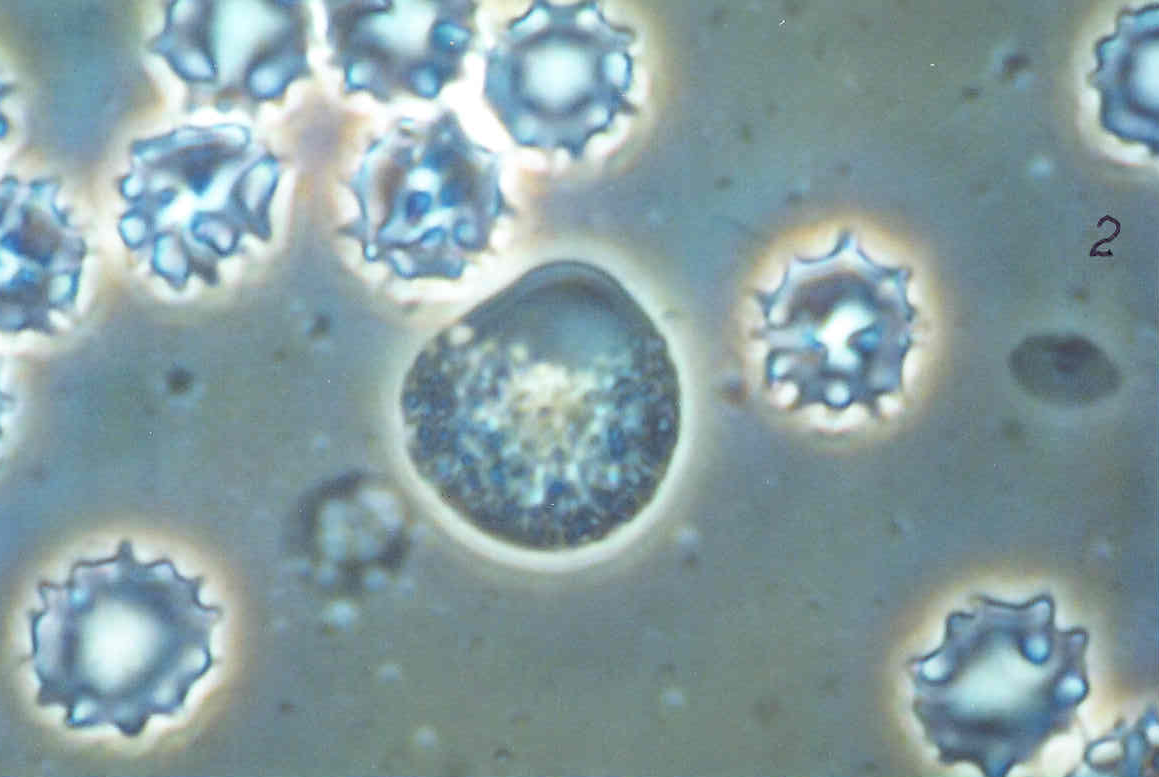
Leukemia - 40x Phase contrast
Leukemia - 40x Phase contrast
Absolutely transparent sample - no structures visible

By increasing the color contrast, all the structures can be seen clearly in their natural colors and with enhanced depth of field.
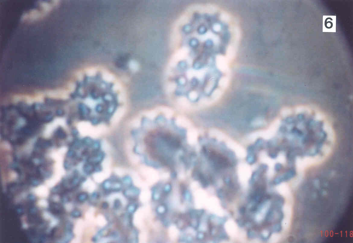
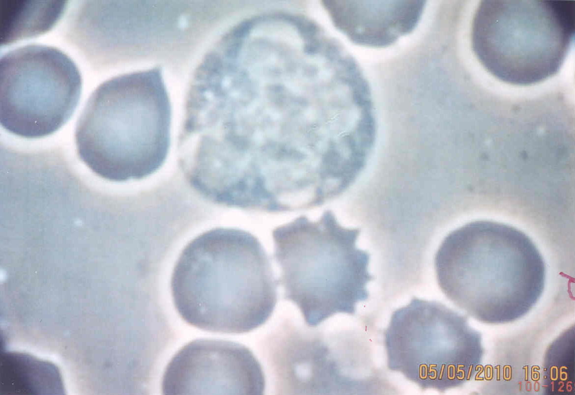
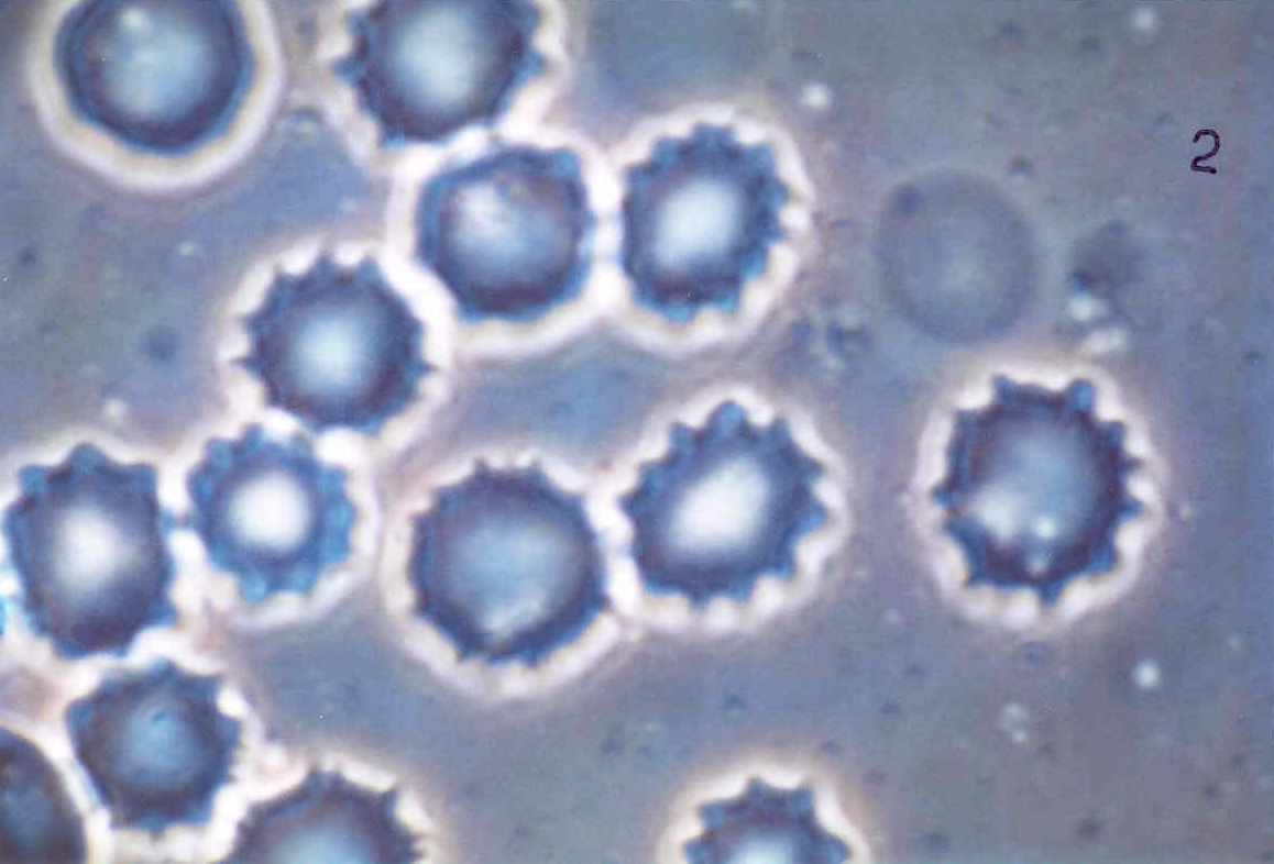
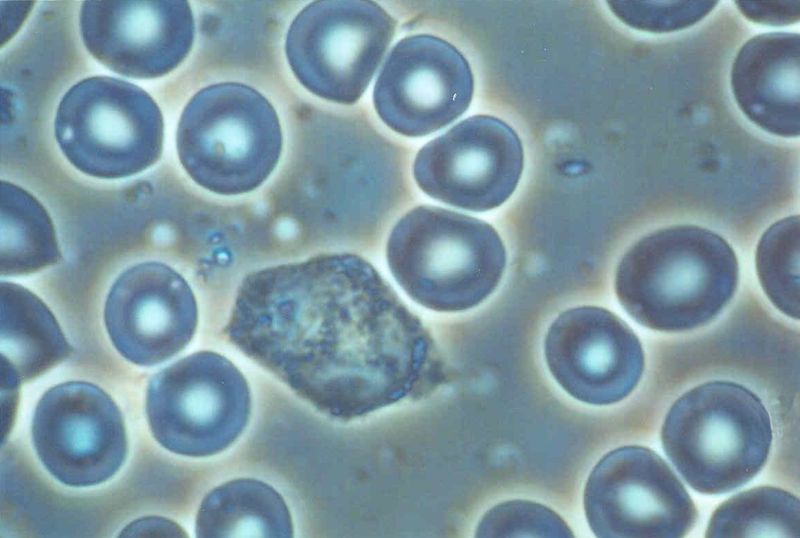
Blood cells heavily infected with cancer - 40x Phase contrast
Blood cells heavily infected with cancer - 40x Phase contrast
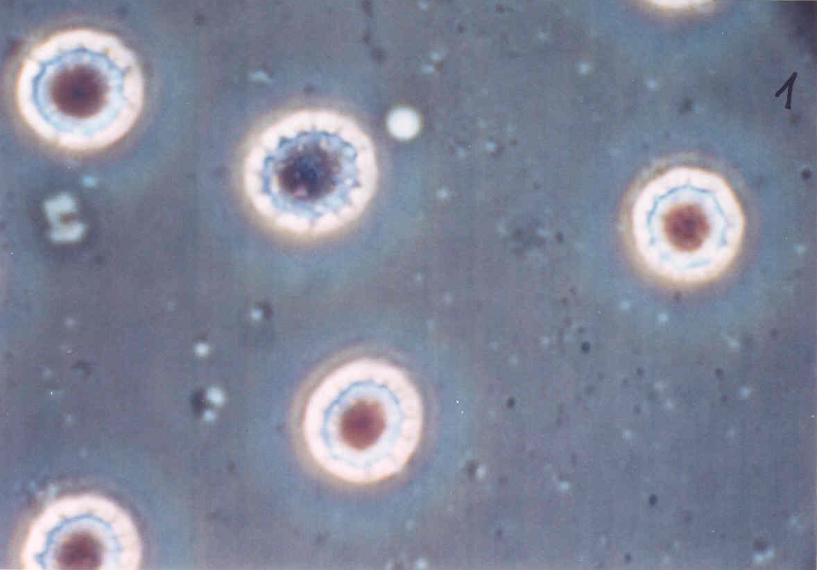
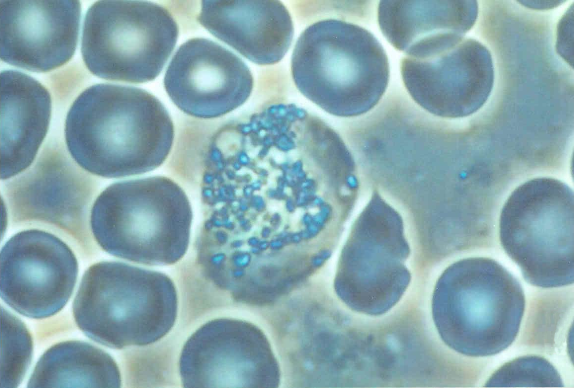
Blood samples 40x Phase contrast - Monocyte / Granulocyte
Blood sample 40x Phase contrast - Macrophage
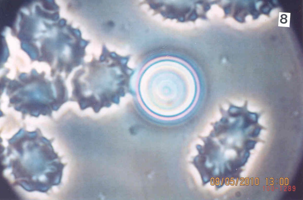
Blood sample 40x Phase contrast - Acanthocyte intracellular infection
Blood sample 40x Phase contrast - possible Rheumatism
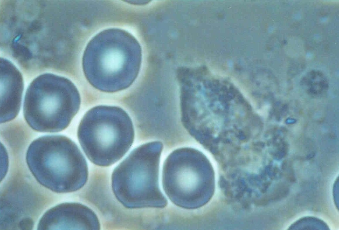
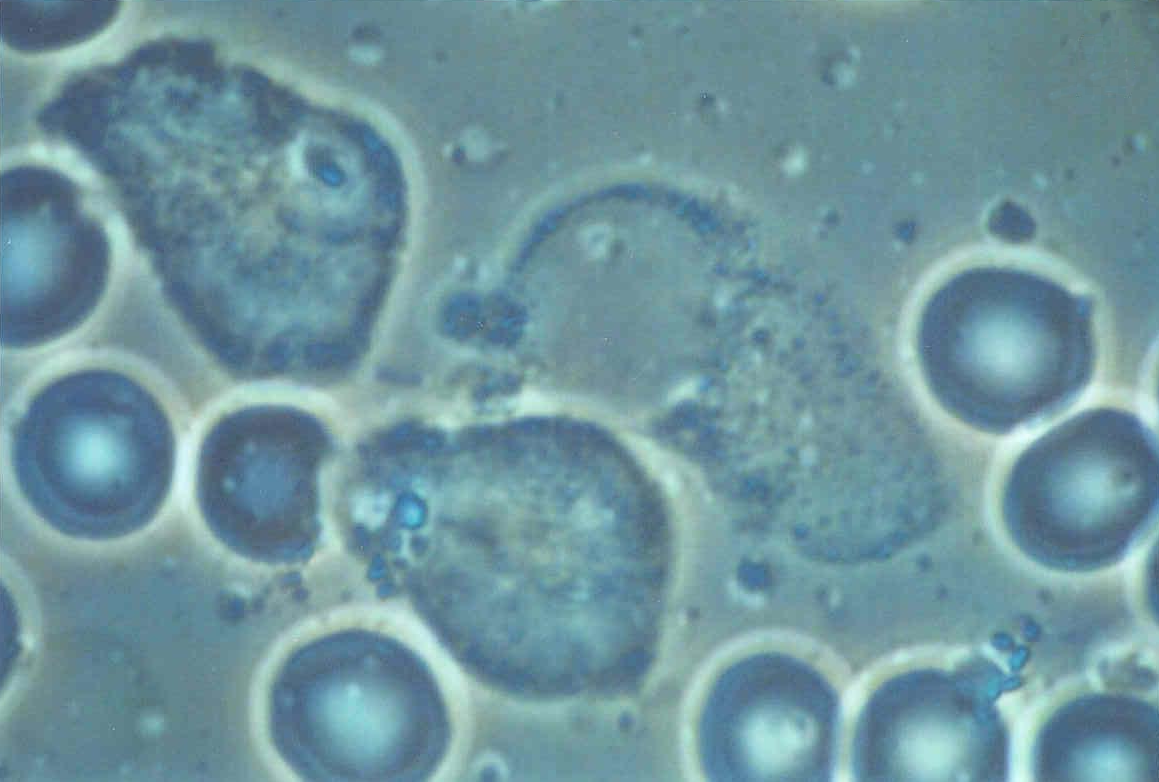
Blood cells heavily infected with cancer
Blood cells heavily infected with cancer
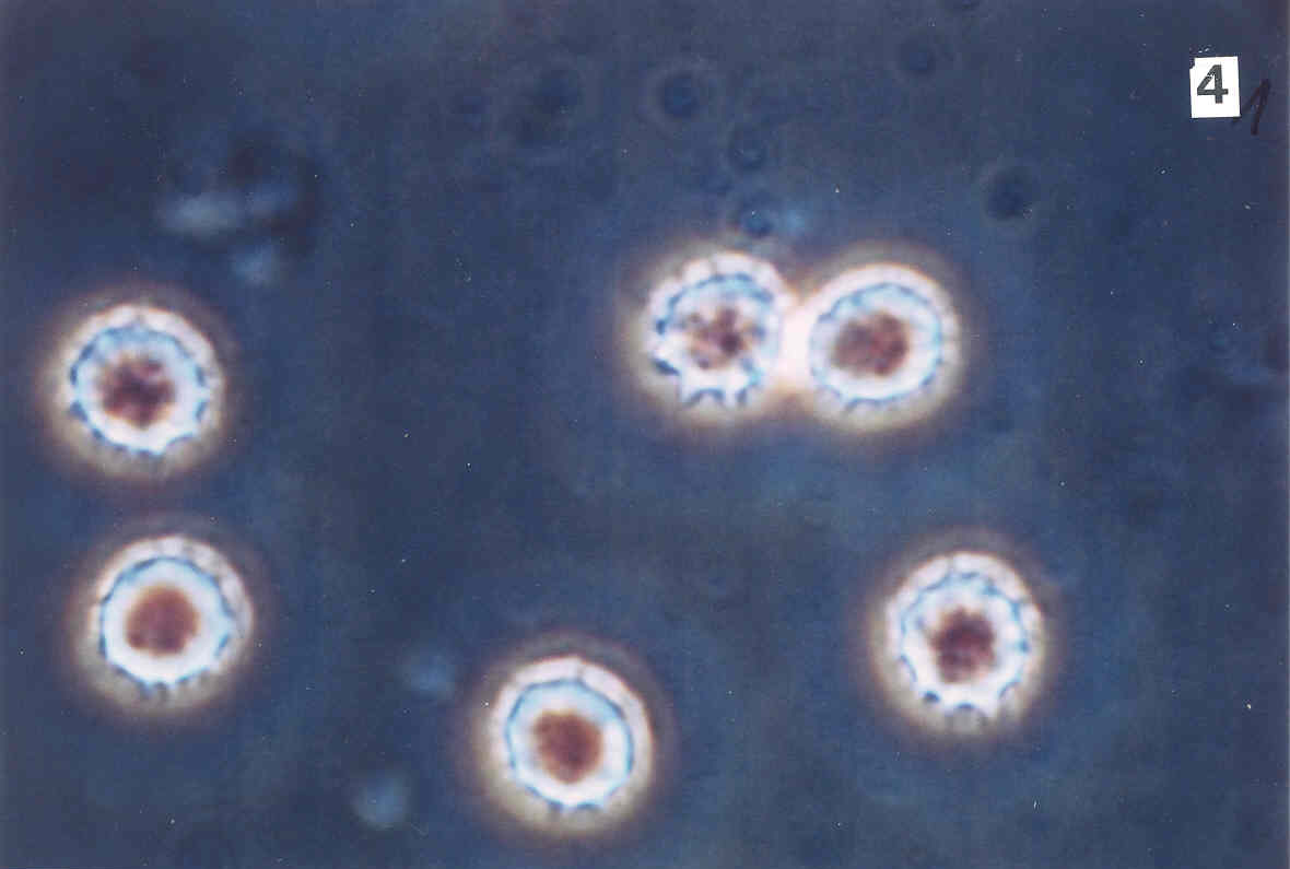
Healthy blood: erythrocytes, granulocytes
Healthy blood: erythrocytes, acanthocytes
Healthy blood: erythrocytes, leucocytes
Healthy blood: erythrocytes, phagocytes


