SeeNano Pro
Products
High Definition Scope in Vivo, in Vitro, in Situ
Ø Operates at room temperature for maximum flexibility
Ø Uses untreated samples to avoid damage and false observations
Ø Non-destructive technique enables living objects to be observed
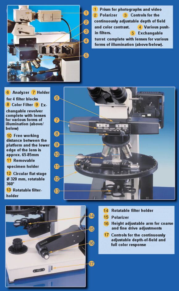
Ø Depth of field is adjustable to penetrate deep samples
Ø Full colour images for detailed analysis
Ø Effective alternative to scanning electron microscope
Ø Resolution and depth of field increased extended
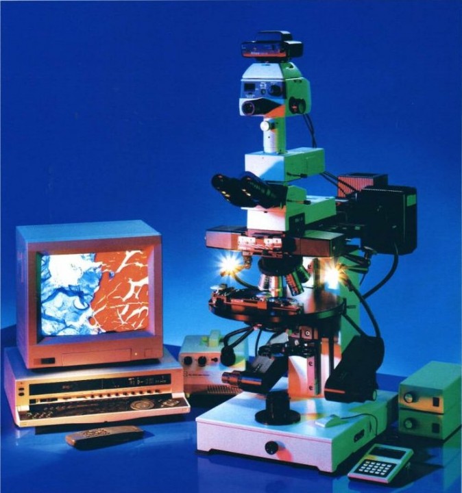
The SeeNano Pro high definition optical microscope provides superior images of cells, tissues, organs and whole preparations in vivo, in vitro and in situ. The major strength of this instrument is its ability to deal with preparations that are difficult with conventional microscopes, such as whole-mounted preparations, living unstained cells, and living organ cultures.
The unique control of illumination and wide range of optical systems gives significantly enhanced flexibility. The SeeNano-Pro surpasses conventional microscopes in its ability to produce superior image quality in difficult applications. It enables the user to control the light beam in order to maximise the image according to the application and the subject.
SeeNano-Pro significantly increases the image quality in terms of detectability, contrast and sharpness of contour. At the same time it sharply reduces internal reflections, double images and flare.
The SeeNano-Pro illumination system has a novel way of controlling the light beam. The result is highly efficient light management that improves the image quality, permits the microscope to penetrate into thick structures and provides for control over depth of focus and depth of field. The combination of penetrating ability with adjustable dept of field represents a unique powerful tool and is a major feature of the SeeNano-Pro.
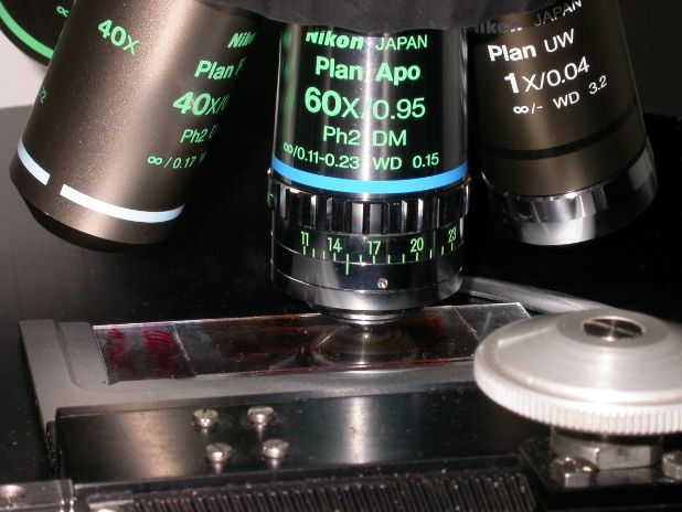
SeeNano-Pro achieves far higher resolution and image quality from dry lenses and long working distances than conventional microscopes. Most remarkable is that the illumination system can employ a condenser lens of low numerical aperture with an objective lens of high "n.a." while maintaining the resolution and image quality expected from the high "n.a." lens. This simply cannot be done with existing microscopes.
In practical terms, this new microscope technology will allow scientists to design and to perform experiments that have not been possible before. It will provide researchers with an important tool for exploring biomedical questions.
Note: Product images should be seen as examples only and are subject to change.

Grayfield Technology
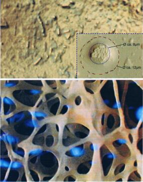
Depth of Field: All our microscopes feature variable depth of field where the true colours and contour sharpness remain clearly discernable, even with ever increasing magnifications comparable with that of mid-range scanning electron microscopes. This makes it possible to vary the depth of field independently of magnification, which also allows much more detail to be seen (see image on right), live and in real time!
No Staining, No Oil: The unique optical system provides so much contrast that staining is not required. This allows you to observe the specimens, under a white light source, in their true (living) state, in vivid contrast and true colours even at the highest magnifications. All objectives remain dry as oil immersion is not required.
Grayfield: An entirely new method in optical microscopy is the Grayfield method. This method allows you to see detailed structures that are otherwise not even visible with conventional phase contrast microscopes. For example, the Grayfield method allows you to observe the in-vitro decomposition processes of blood. During this transitional phase new viruses and structures arise, which tend to decay and could previously not be made visible due to the lack of suitable microscope techniques.