SeeNano versus Fluorescence Microscope
Comparison
Fluorescence microscopes are often used due to the generally better image quality when compared to white light microscopes. This is based on the use on ultra-violet light with its shorter wavelengths therefore allowing a better resolution.
Although the previous range of microscopes could only use white light, the SeeNano can also make use of Fluorescence where needed.
When using the Grayfield Lens System, the white light source used on the SeeNano provides a better more detailed image than UV can do, without the inherent dangers associated with UV light usage.
Fluorescence Microscope
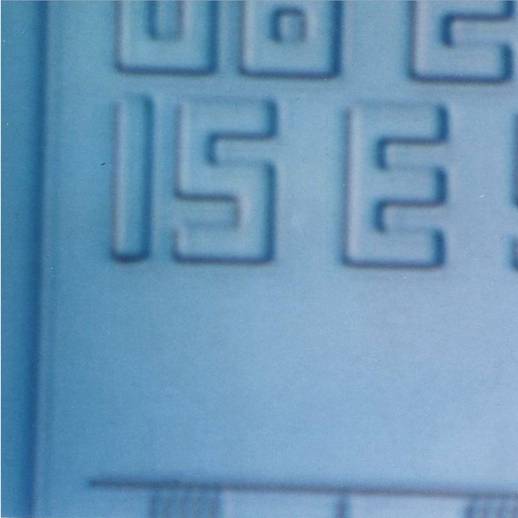
Part of a computer chip, that has been treated for viewing under a Scanning Electron Microscope (SEM), as seen with UV light.
SeeNano Microscope
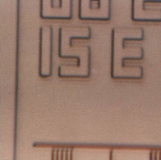
The same chip, now seen under white light using the SeeNano microscope. The white light image provides a clearer image.
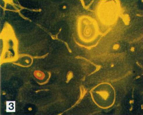
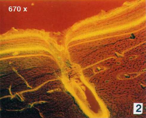
Cross section of a bone from a racing horse:
Fluorescence Microscope

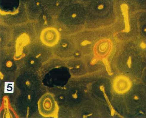
SeeNano Microscope
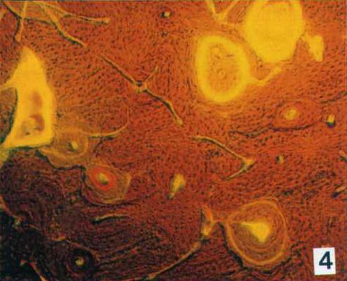
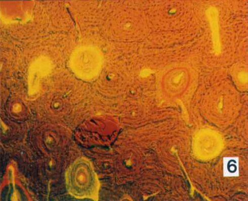
The images on the left show the normal fluorescence images, with minimised over exposure. Normal fluorescence images cannot show the structures beneath them at the same time. In order to make the images on the right, a combination of fluoresence and phase contrast was used to obtain an image with the detailed background.