Fluorescence Tagging - New 2015
Technology
The new SeeNano range of microscopes introduces a new capabilitiy not available in our previous range - Fluorescence Tagging. On this page, we will give you a sample of what we can achieve with this new feature in combination with our Grayfield contrast system.
Original Samples from Riley/IUPUI
Centrosome Reference Pictures
First Attempt at Fluorescence Tagging
(Click on images to enlarge)
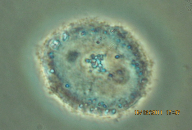
White light stimulus - unaltered image, magnification ≈ 3,000x
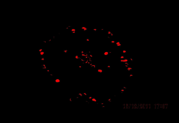
Computer extracted image - White Light stimulus only
No filters - magnification ≈ 3,000x
No filters - magnification ≈ 3,000x
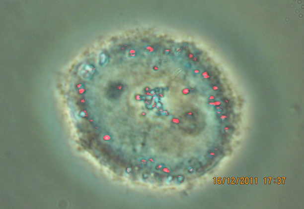
Composite of White Light + Computer generated image,
magnification ≈ 3,000x
magnification ≈ 3,000x
Using Excitation and Emission Filters
To confirm fluorescence imaging
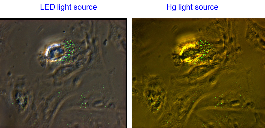
The SeeNano prototype was modified to add fluorescence capability:
- Added Fluorescence filter holders for Excitation and Emission filtering
- Added a Mercury Light Source for fluorescence stimulation, made switchable with the LED Light source used for white light illumination
- Received considerable support regarding procedures and critical considerations from the University of Hamburg, who also provided fluorophore infused live cell samples for experimentation
Samples from University of Hamburg were Peroxisomes Infused with GFP fluorophores.
The filters used on the SeeNano Prototype were:
- Excitation: Edmund Optics 51022x EGFP
- Emmission: Edmund Optics 51022m EGFP
Nikon Image of Peroxisome Sample
Imaged using: Nikon A1 Confocal Laser System, provided by University of Hamburg
(Click on images to enlarge)
DIC (Raw image)
Magnification ≈ 1,000x
Magnification ≈ 1,000x
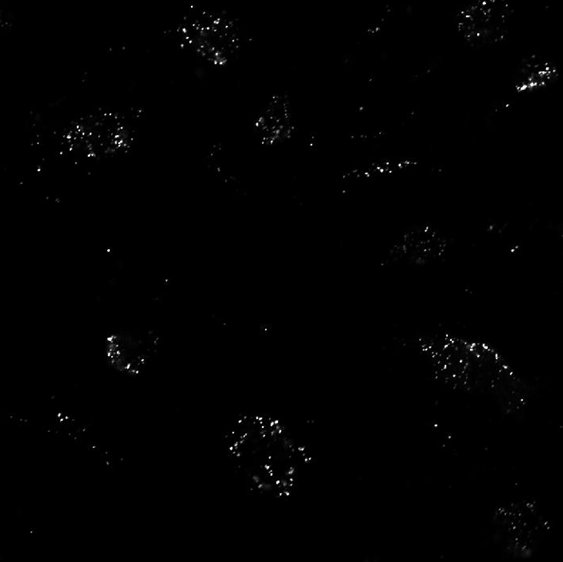
Fluorescence scanned image
Magnification ≈ 1,000x
Magnification ≈ 1,000x
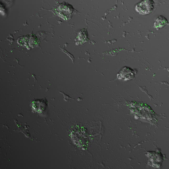
Composite processed image
Magnification ≈ 1,000x
Magnification ≈ 1,000x
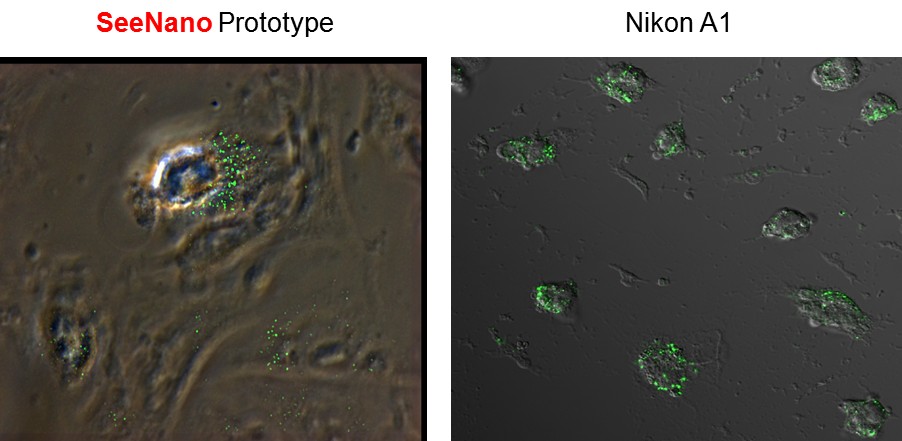
SeeNano Image of Peroxisome Sample
Imaged using: SeeNano prototype Grayfield microscope System
(Click on images to enlarge)
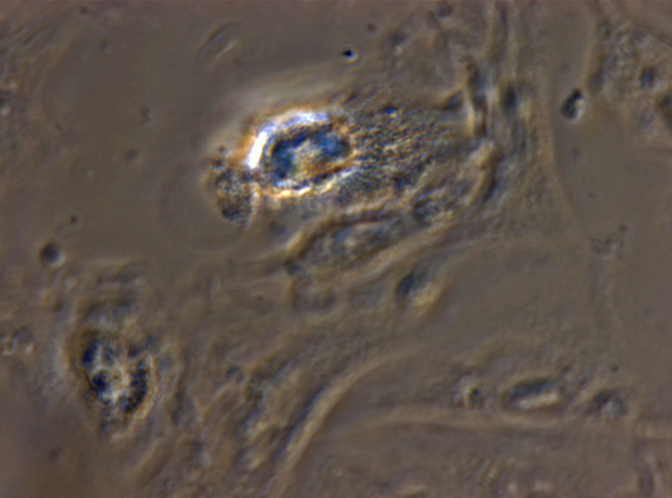
Phase Contrast (raw image)
Transmission Mode, Light source: WL=LED, Wide Field Capture
Magnification ≈ 2,500x
Transmission Mode, Light source: WL=LED, Wide Field Capture
Magnification ≈ 2,500x
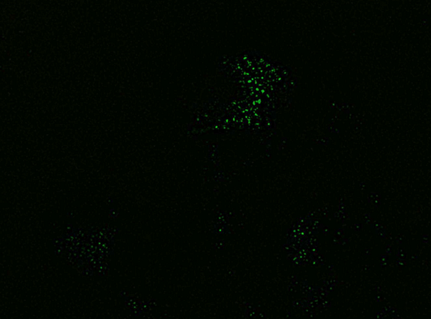
Fluorescence processed image
Transmission Mode, Light source: FL=Hg, Wide Field Capture
Magnification ≈ 2,500x
Transmission Mode, Light source: FL=Hg, Wide Field Capture
Magnification ≈ 2,500x
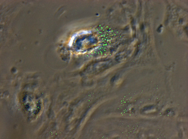
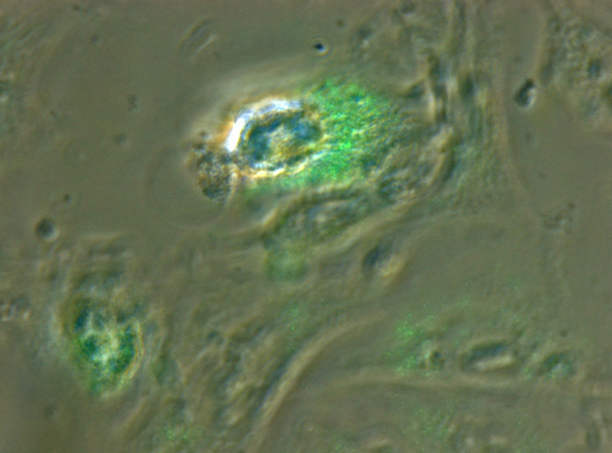
Composite Raw PC+Enhanced Fluorescence
Transmission Mode, Light source: FL=Hg and WL=LED, Wide Field Capture
Magnification ≈ 2,500x
Transmission Mode, Light source: FL=Hg and WL=LED, Wide Field Capture
Magnification ≈ 2,500x
Phase Contrast (raw image)
Transmission Mode, Light source: WL=LED, Wide Field Capture
Magnification ≈ 2,500x
Transmission Mode, Light source: WL=LED, Wide Field Capture
Magnification ≈ 2,500x
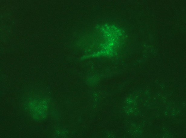
Fluorescence (raw image)
Transmission Mode, Light source: FL=Hg, Wide Field Capture
Magnification ≈ 2,500x
Transmission Mode, Light source: FL=Hg, Wide Field Capture
Magnification ≈ 2,500x
Composite raw fluorescence + white light
Transmission Mode, Light source: FL=Hg, WL=LED, Wide Field Capture
Magnification ≈ 2,500x
Transmission Mode, Light source: FL=Hg, WL=LED, Wide Field Capture
Magnification ≈ 2,500x