SeeNano Lab
Darkfield was Yesterday..., See more with Grayfield and in Color!
No more Staining or oil immersion required!
Variable extended depth of field!
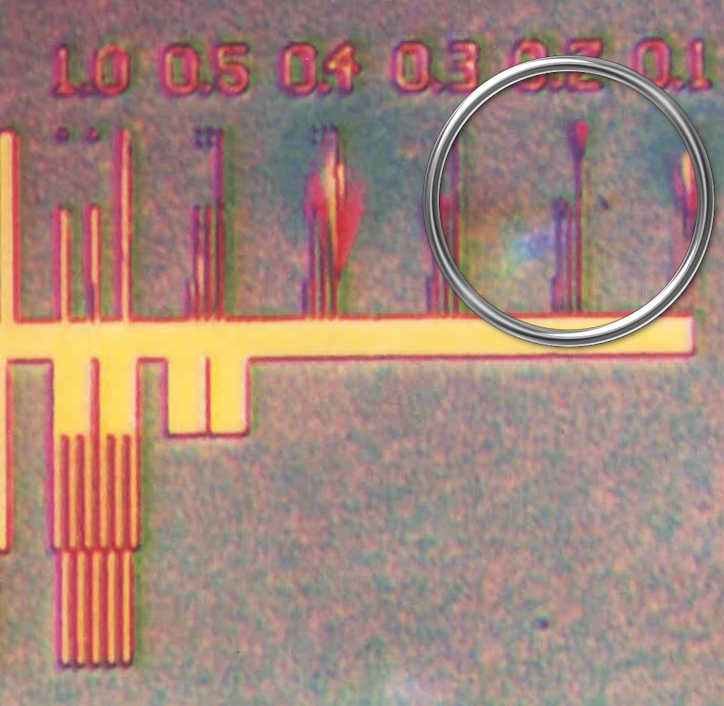
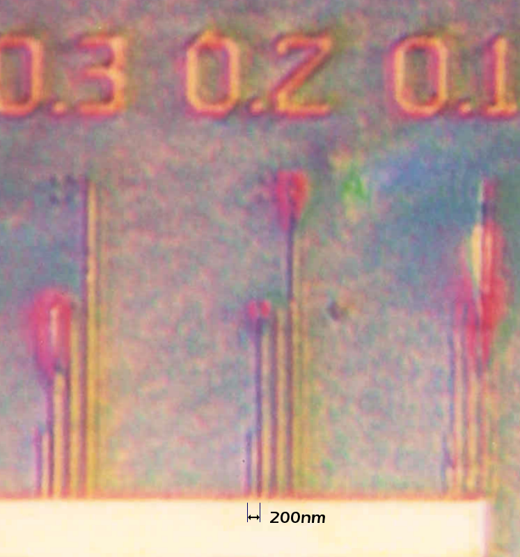
Practical resolution: 200nm in natural colors
Variable color contrast even for transparent samples.
Our proprietary Grayfield contrast method gives you full contrast images even at high magnifications
No staining
No messy oil
Full contrast
Full natural colors
Living samples
Affordable price
Click on the images to enlarge...
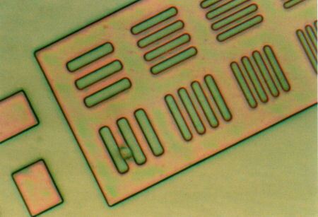
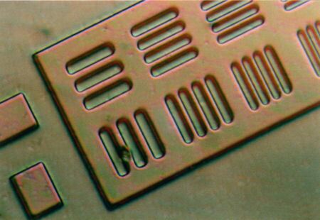
Variable Depth of Field
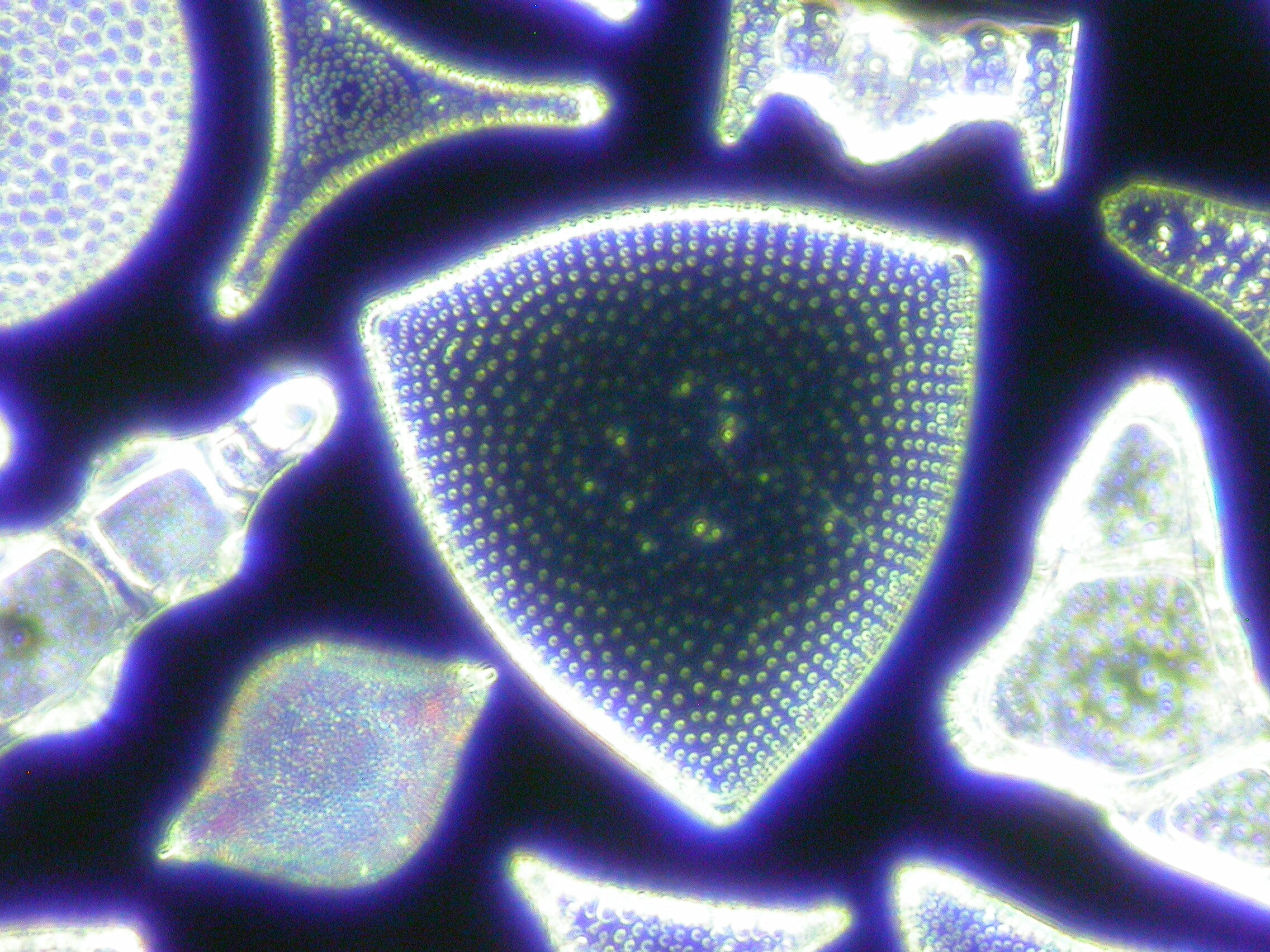
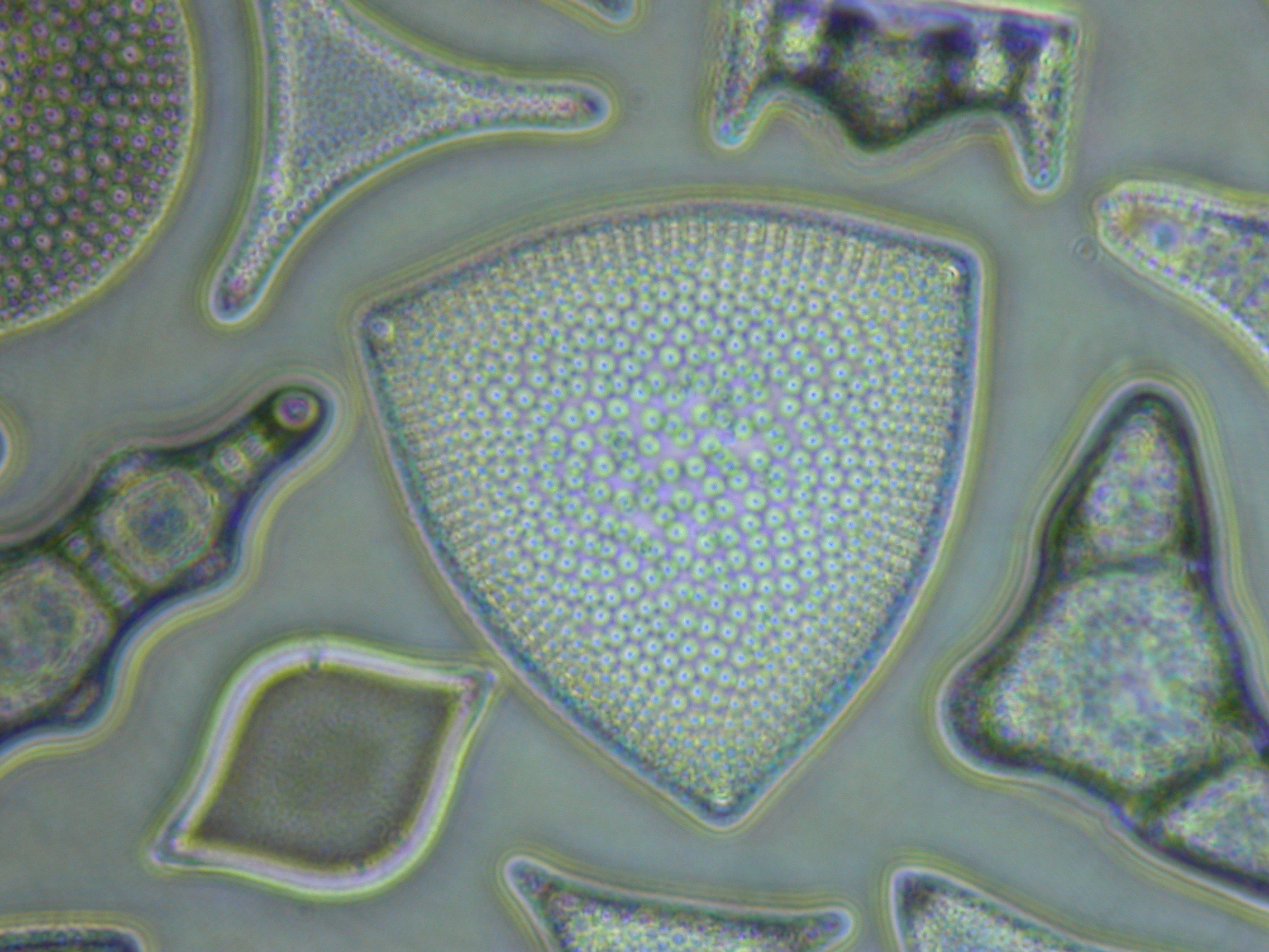
Transparent Samples
Click on the images to enlarge...
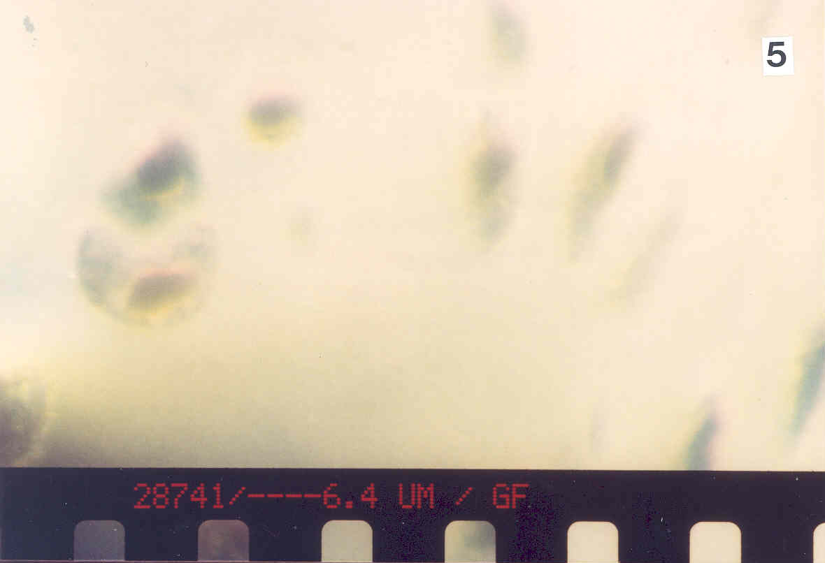

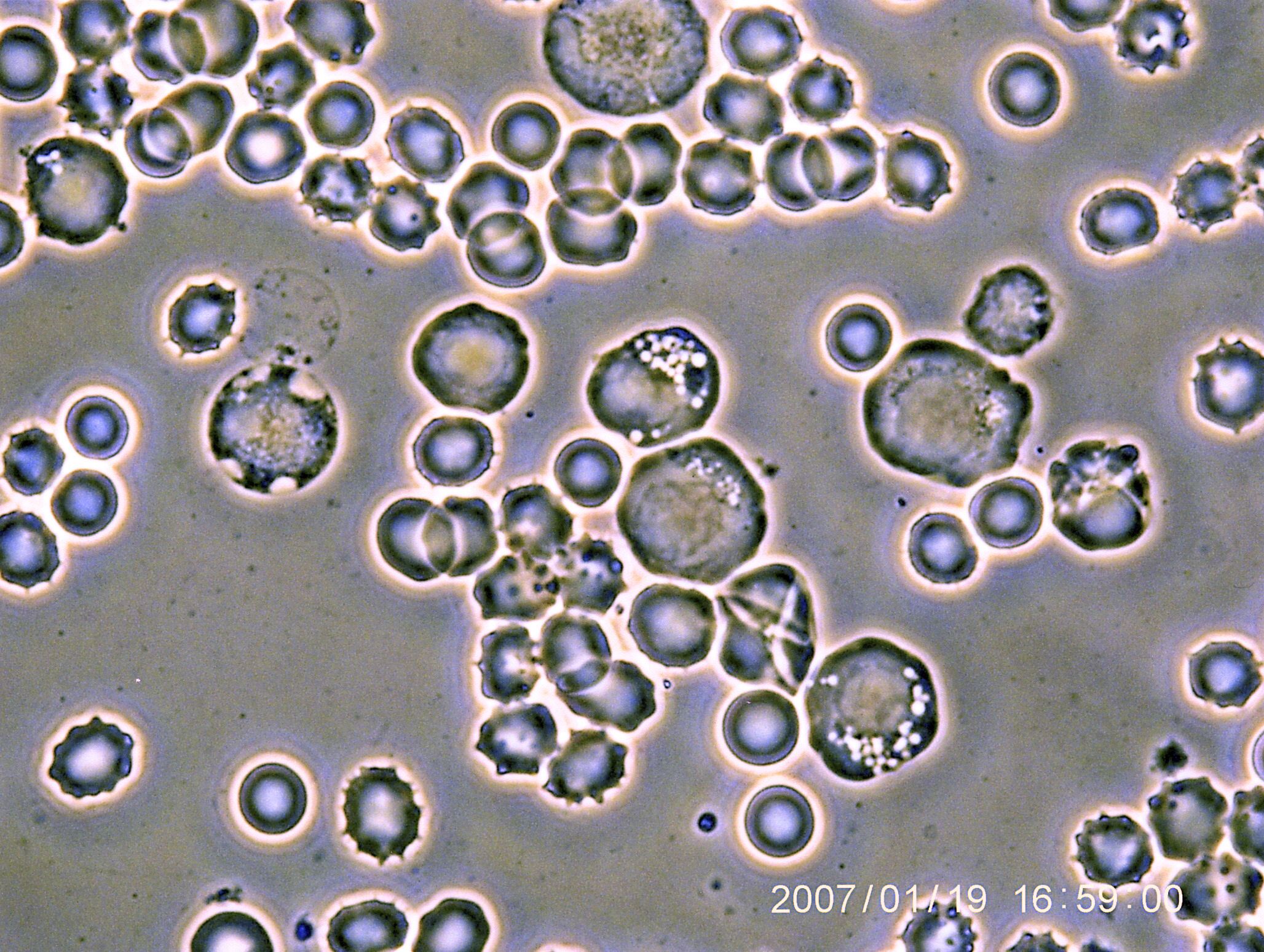
Blood Samples
(Transmitted light only)
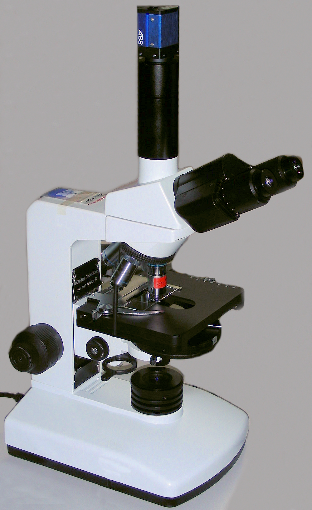
5x turret equipped with three standard objectives for transmitted light:phase contrast (enhanced)
40x darkfield (improved), 60x transmitted/grayfield
Optional 80x grayfield objective, NA 0.9 (extreme depth of field, etc.)
4x special condensers for a wide range of applications
Illumination system for transmitted light (e.g.biological applications)
True resolution up to 200nm with white light source
True magnification possible up to at 4500x
Height adjustable stage
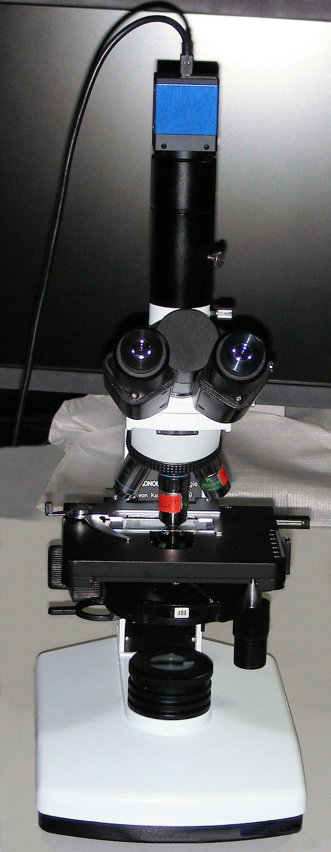
Binocular viewing head with C-mount port for attaching photo and video cameras
20x Ocular lens
Very stable base
Made in Germany
Ideal for biological and medical applications with high demands. The turret is fitted with high quality objectives and a condensor. e.g. for our enhanced positive and negative phase contrast system for brightfield, darkfield and grayfield use.
Staining and oil immersion are not required which is ideal for live cell and blood analysis in their natural colors and in real time.
Full color high contrast images are possible even up to the full resolution of 200nm and variable depth of field allows you to see the images with a sharpness and depth not possible with any other optical microscopes.
Click here to see more sample images...
(Incident light only)
5x turret equipped with three standard objectives for incident light:phase contrast (enhanced)
Range of optional objectives to meet your applications, NA 0.9 (extreme depth of field, etc.)
Illumination system for incident light
Ideal for imaging solid objects like electronic components, metallurgy, plastics, etc.
True resolution up to 200nm with white light source
True magnification possible up to at 4500x
Height adjustable stage
Binocular viewing head with C-mount port for attaching photo and video cameras
20x Ocular lens
Very stable base
Made in Germany
Ideal for applications for imaging solid objects in high resolution and in real time. e.g. electronic components, metallurgy, plastics, etc. up to a resolution of 200nm.
Staining and oil immersion are not required which is ideal for imaging computer components, etc. as they are without any added coatings or contrast agents of any kind (remains dry). Unlike other laser scanning based systems, these microscopes work purely optically in real time without the need for computer image processing, etc.
Full color high contrast images are possible even up to the full resolution of 200nm and variable depth of field allows you to see the images with a sharpness and depth not possible with any other optical microscopes.
Click here to see more sample images...
Note: For technical reasons, it is not feasible to build a SeeNano Lab for both Transmitted and Incident light.
Grayfield Technology